Introduction
Pneumonia is a serious condition that affects the lungs. It can be caused by bacteria, viruses, or fungi and can lead to breathing difficulties, chest pain, fever, and other severe symptoms. Diagnosing pneumonia quickly and accurately is crucial, as it can help prevent complications and improve outcomes. In this article, we’ll explore the major symptoms of pneumonia, the physical examination that healthcare providers use to diagnose it, imaging techniques such as X-rays, and the various blood tests that are used. We’ll also discuss causes and risk factors and provide tips from real-life case studies to help readers understand how diagnosis works in practice.
Symptoms Checklist
The symptoms of pneumonia can vary widely in their severity and duration, and not everyone will experience all of them. Here are some of the most common symptoms:
- Coughing
- Chest pain
- Shortness of breath
- Fever
- Fatigue
- Sweating and shaking
- Loss of appetite
- Headache
- Confusion (especially in older adults)
- Bluish lips or nails (due to lack of oxygen)
If you experience any of these symptoms, it’s important to take action. Keep track of your symptoms and their severity, and contact your healthcare provider if they worsen or if you have trouble breathing or maintaining normal oxygen levels.
Physical Examination
In addition to asking you about your symptoms and medical history, your healthcare provider will perform a physical exam to check for signs of pneumonia. These exams often include listening to your lungs with a stethoscope, checking your body temperature, and assessing the color of your skin and fingernails. Your healthcare provider will also check for any chest pain, coughing, or difficulty breathing, which could indicate pneumonia.
During the lung examination, healthcare providers will use the stethoscope to listen to your breathing. They are usually listening for “crackling” or “rattling” sounds, which suggest fluid or mucus in the lungs. They may also listen for a “whistling” sound called wheezing, which indicates difficulty breathing or asthma.
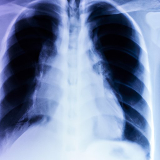
X-Ray Explanation
X-rays are often used to diagnose pneumonia. The process involves standing close to a large machine and holding still for a few seconds while special images of the chest are taken. These images can help doctors see if there is any inflammation, fluid buildup, or other signs of infection in the lungs.
X-rays can help to determine the location and extent of the infection, and can be used to monitor the condition over time. They also help to differentiate pneumonia from other lung conditions, such as bronchitis or asthma. If an X-ray does not provide enough information, other imaging tests like CT scans may be performed.
Causes and Risk Factors
Pneumonia can be caused by a variety of factors, with different risk factors that may influence the diagnosis. Common risk factors include:
- Age (especially infants, young children, and older adults)
- Smoking or exposure to secondhand smoke
- Chronic obstructive pulmonary disease (COPD)
- Diabetes
- Weakened immune system (due to chemotherapy, steroids, or HIV/AIDS)
- Recent hospitalization
- Exposure to certain chemicals or pollutants
If you have any of these risk factors, it’s important to be vigilant for signs of pneumonia and to follow recommendations from your healthcare providers in order to help prevent and manage the condition.
Blood Tests
In addition to physical exams and imaging, blood tests may be performed to diagnose pneumonia. These tests can measure white blood cell counts, blood oxygen levels, and other markers of infection.
White blood cells are part of the body’s immune system, responsible for fighting off bacteria and other infections. If levels are abnormal, it could indicate an infection like pneumonia. Blood oxygen levels can also be checked to ensure that the lungs are functioning properly. Other markers such as C-reactive protein (CRP) can help indicate the severity of the infection.
Case Studies
To understand how diagnosis works in practice, let’s look at some real-life case studies.
Case One: A 57-year-old man with a history of smoking presents to his primary care provider with a cough that has persisted for over a week. He also reports chest pain and fever. After taking his medical history and performing a physical exam, the healthcare provider orders a chest X-ray and blood tests. The tests show that the man has pneumonia, and he is prescribed antibiotics.
Case Two: An 80-year-old woman is brought to the emergency department by her daughter, who reports that her mother has been increasingly confused and lethargic over the past several days. The healthcare providers notice that her breathing is labored, and her oxygen levels are low. After performing a physical exam and ordering blood tests and a chest X-ray, they diagnose her with pneumonia caused by a bacterial infection. She is admitted to the hospital and given antibiotics and supportive care, and gradually improves over time.
Conclusion
In conclusion, pneumonia is a serious illness that requires prompt and accurate diagnosis. Recognizing and monitoring common symptoms, understanding physical exams and imaging, and interpreting blood test results are all essential in diagnosing pneumonia accurately. The causes and risk factors of pneumonia can also contribute to the diagnosis. It’s important to consult with healthcare providers if you suspect that you have pneumonia, as early detection and treatment can lead to better outcomes. Seeking information and resources can also help readers learn more about prevention and treatment options that can help prevent complications related to pneumonia.